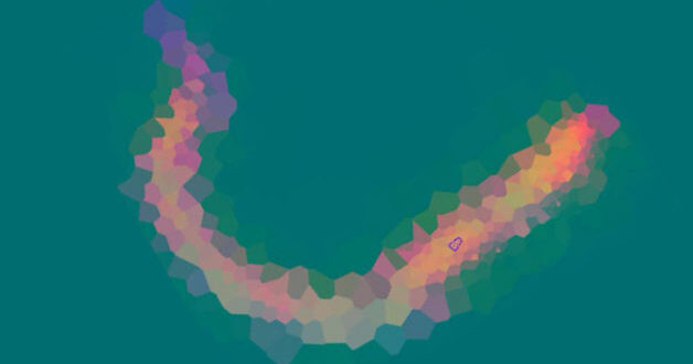
Example of the tiling pattern used in scanning a C. elegans roundworm. The non-grid-based pattern gives the sampling algorithm greater flexibility to quickly zero in on areas of interest. (Credit: Elizabeth Holman/Caltech and Yuan-Sheng Fang/Berkeley Lab)
Question: What do a roundworm, a Sharpie pen, and high-vacuum grease have in common? Answer: They’ve all been analyzed in recent proof-of-principle microscopy experiments at Berkeley Lab’s Advanced Light Source (ALS).
In the journal Communications Biology, researchers from Caltech, UC Berkeley, and the Berkeley Synchrotron Infrared Structural Biology Imaging Program (BSISB) reported a more efficient way to collect “high-dimensional” infrared images – where each pixel contains rich physical and chemical information. With the new method, scans that would’ve taken up to 10 hours to complete can now be done in under an hour, potentially broadening the scope of biological spectromicroscopy to time-sensitive experiments.
“We realized that sampling our model organism – the small roundworm C. elegans – as it changes over time was challenging for software rather than hardware reasons,” said Elizabeth Holman, a graduate student in chemistry at Caltech and co-first author of the paper. “For example, image sampling was limited to uniform-grid raster scans with rectangular boundaries and fixed distances between sample points.”
The new technique, implemented at the ALS with co-first author Yuan-Sheng Fang, a graduate student in physics at UC Berkeley, uses a grid-less, adaptive approach that autonomously increases sampling in areas displaying greater physical or chemical contrast. In the proof-of-concept infrared microscopy experiments, the researchers examined two samples.
The first was a two-component system in which both components (permanent-marker ink and high-vacuum grease) were well characterized. Details of the sample were very difficult to see clearly with the naked eye, so it was a good test of how the software would perform with minimal guidance from a human experimenter. The second sample was a live, larval-stage C. elegans, a biological model system studied by thousands of researchers.
In both cases, autonomous adaptive data acquisition (AADA) methods clearly outperformed nonadaptive methods. In the second example, increased sampling density corresponded with known C. elegans anatomical features, and the head region was mapped in 45 minutes versus about 4.9 hours using commercially available software.
“Outside of our specific published work, the results suggest that integrating AADA into existing scanning-based satellite, drone, and/or microscope techniques can facilitate research in fields ranging from hyperspectral remote sensing to ocean and space exploration,” said Holman.
You just read:
EIN Presswire’s priority is source transparency. We do not allow opaque clients, and our editors try to be careful about weeding out false and misleading content.
As a user, if you see something we have missed, please do bring it to our attention. Your help is welcome. EIN Presswire, Everyone’s Internet News Presswire™,
tries to define some of the boundaries that are reasonable in today’s world. Please see our
Editorial Guidelines
for more information.
Submit your press release